

Skogen, K;
Schulz, A;
Helseth, E;
Ganeshan, B;
Dormagen, JB;
Server, A;
(2019)
Texture analysis on diffusion tensor imaging: discriminating glioblastoma from single brain metastasis.
Acta Radiologica
, 60
(3)
pp. 356-366.
10.1177/0284185118780889.

Preview |
Text (Accepted manuscript)
Ganeshan_Texture analysis on diffusion tensor imaging.pdf - Accepted Version Download (443kB) | Preview |
![[thumbnail of Figure 1]](https://discovery.ucl.ac.uk/10054586/3/Texture%20analysis%20Figure%201.jpg)  Preview |
Image (Figure 1)
Texture analysis Figure 1.jpg - Accepted Version Download (1MB) | Preview |
![[thumbnail of Figure 2]](https://discovery.ucl.ac.uk/10054586/8/Texture%20analysis%20Figure%202.jpg)  Preview |
Image (Figure 2)
Texture analysis Figure 2.jpg - Accepted Version Download (3MB) | Preview |
![[thumbnail of Figure 3]](https://discovery.ucl.ac.uk/10054586/13/Texture%20analysis%20Figure%203.jpg)  Preview |
Image (Figure 3)
Texture analysis Figure 3.jpg - Accepted Version Download (122kB) | Preview |
![[thumbnail of Figure 4]](https://discovery.ucl.ac.uk/10054586/18/Texture%20analysis%20Figure%204.jpg)  Preview |
Image (Figure 4)
Texture analysis Figure 4.jpg - Accepted Version Download (261kB) | Preview |
![[thumbnail of Figure 5]](https://discovery.ucl.ac.uk/10054586/23/Texture%20analysis%20Figure%205.jpg)  Preview |
Image (Figure 5)
Texture analysis Figure 5.jpg - Accepted Version Download (1MB) | Preview |
Abstract
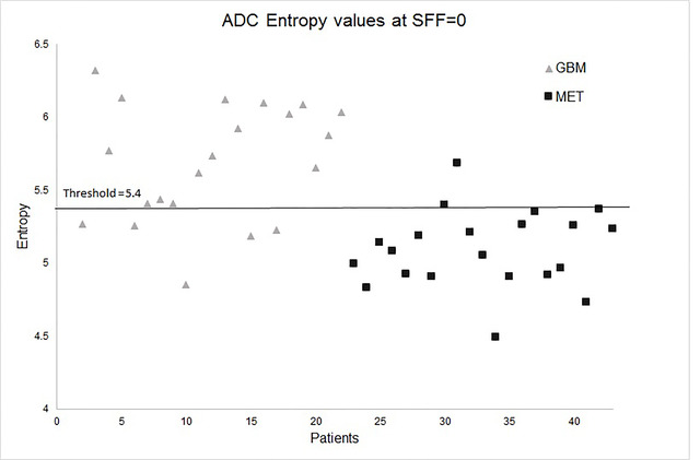
BACKGROUND: Texture analysis has been done on several radiological modalities to stage, differentiate, and predict prognosis in many oncologic tumors. PURPOSE: To determine the diagnostic accuracy of discriminating glioblastoma (GBM) from single brain metastasis (MET) by assessing the heterogeneity of both the solid tumor and the peritumoral edema with magnetic resonance imaging (MRI) texture analysis (MRTA). MATERIAL AND METHODS: Preoperative MRI examinations done on a 3-T scanner of 43 patients were included: 22 GBM and 21 MET. MRTA was performed on diffusion tensor imaging (DTI) in a representative region of interest (ROI). The MRTA was assessed using a commercially available research software program (TexRAD) which applies a filtration histogram technique for characterizing tumor and peritumoral heterogeneity. The filtration step selectively filters and extracts texture features at different anatomical scales varying from 2 mm (fine) to 6 mm (coarse). Heterogeneity quantification was obtained by the statistical parameter entropy. A threshold value to differentiate GBM from MET with sensitivity and specificity was calculated by receiver operating characteristic (ROC) analysis. RESULTS: Quantifying the heterogeneity of the solid part of the tumor showed no significant difference between GBM and MET. However, the heterogeneity of the GBMs peritumoral edema was significantly higher than the edema surrounding MET, differentiating them with a sensitivity of 80% and specificity of 90%. CONCLUSION: Assessing the peritumoral heterogeneity can increase the radiological diagnostic accuracy when discriminating GBM and MET. This will facilitate the medical staging and optimize the planning for surgical resection of the tumor and postoperative management.
| Type: | Article |
|---|---|
| Title: | Texture analysis on diffusion tensor imaging: discriminating glioblastoma from single brain metastasis |
| Location: | England |
| Open access status: | An open access version is available from UCL Discovery |
| DOI: | 10.1177/0284185118780889 |
| Publisher version: | https://doi.org/10.1177%2F0284185118780889 |
| Language: | English |
| Additional information: | This version is the author accepted manuscript. For information on re-use, please refer to the publisher’s terms and conditions. |
| Keywords: | Glioblastoma, brain metastases, diffusion tensor imaging, magnetic resonance imaging, peritumoral edema, texture analysis |
| UCL classification: | UCL UCL > Provost and Vice Provost Offices > School of Life and Medical Sciences UCL > Provost and Vice Provost Offices > School of Life and Medical Sciences > Faculty of Medical Sciences UCL > Provost and Vice Provost Offices > School of Life and Medical Sciences > Faculty of Medical Sciences > Div of Medicine UCL > Provost and Vice Provost Offices > School of Life and Medical Sciences > Faculty of Medical Sciences > Div of Medicine > Department of Imaging |
| URI: | https://discovery.ucl.ac.uk/id/eprint/10054586 |

Archive Staff Only
 |
View Item |