

Mitchison, HM;
Shoemark, A;
(2017)
Motile cilia defects in diseases other than primary ciliary dyskinesia: The contemporary diagnostic and research role for transmission electron microscopy.
Ultrastructural Pathology
, 41
(6)
pp. 415-427.
10.1080/01913123.2017.1370050.

Preview |
Text
Mitchison_Shoemark revised text.pdf - Accepted Version Download (484kB) | Preview |
Preview |
Text
Mitchison_Table 1. Organ involvement in ciliopathies.pdf - Accepted Version Download (248kB) | Preview |
![[thumbnail of Mitchison_Figure 1.jpg]](https://discovery.ucl.ac.uk/10044257/11/Mitchison_Figure%201.jpg)  Preview |
Image
Mitchison_Figure 1.jpg - Accepted Version Download (208kB) | Preview |
![[thumbnail of Mitchison_Figure 2.jpg]](https://discovery.ucl.ac.uk/10044257/16/Mitchison_Figure%202.jpg)  Preview |
Image
Mitchison_Figure 2.jpg - Accepted Version Download (235kB) | Preview |
Abstract
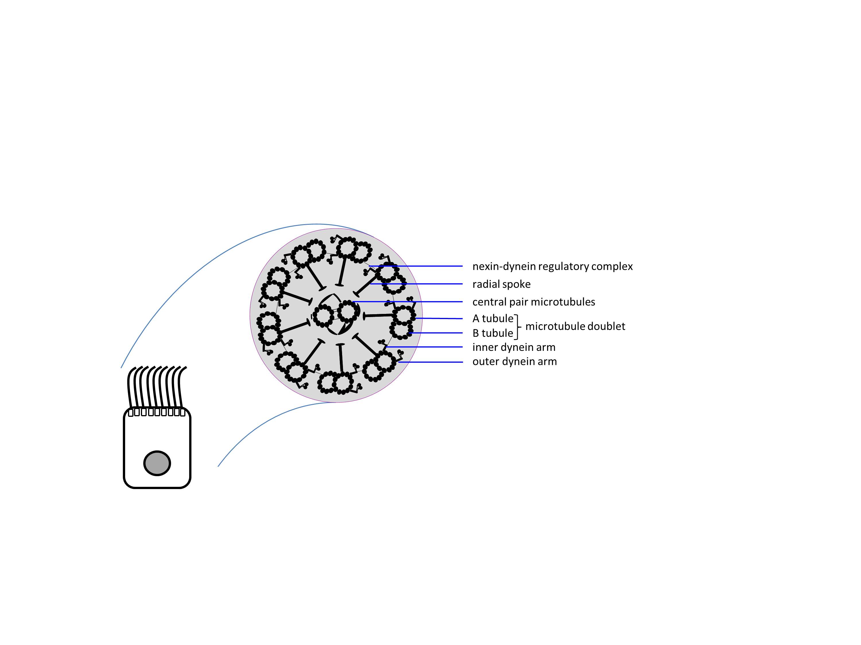
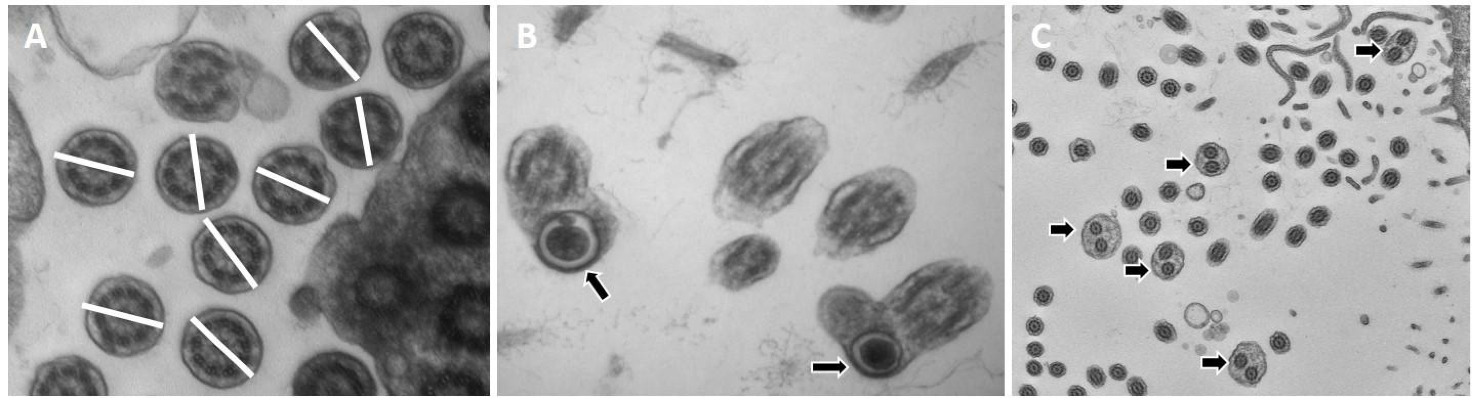
Ultrastructural studies have underpinned the cell biological and clinical investigations of the varied roles of motile cilia in health and disease, with a long history since the 1950s. Recent developments from transmission electron microscopy (TEM; cryo-electron microscopy, electron tomography) have yielded higher resolution and fresh insights into the structure and function of these complex organelles. Microscopy in ciliated organisms, disease models, and in patients with ciliopathy diseases has dramatically expanded our understanding of the ubiquity, multisystem involvement, and importance of cilia in normal human development. Here, we review the importance of motile cilia ultrastructural studies in understanding the basis of diseases other than primary ciliary dyskinesia.
| Type: | Article |
|---|---|
| Title: | Motile cilia defects in diseases other than primary ciliary dyskinesia: The contemporary diagnostic and research role for transmission electron microscopy |
| Open access status: | An open access version is available from UCL Discovery |
| DOI: | 10.1080/01913123.2017.1370050 |
| Publisher version: | http://doi.org/10.1080/01913123.2017.1370050 |
| Language: | English |
| Additional information: | © 2017 Taylor & Francis. This version is the author accepted manuscript. For information on re-use, please refer to the publisher’s terms and conditions. |
| Keywords: | Science & Technology, Technology, Life Sciences & Biomedicine, Microscopy, Pathology, Transmission electron microscopy, ciliopathy disease, non-respiratory, motile cilia, RIGHT AXIS DETERMINATION, DYNEIN REGULATORY COMPLEX, CONGENITAL HEART-DISEASE, HUMAN FALLOPIAN-TUBES, LEFT-RIGHT ASYMMETRY, RESPIRATORY CILIA, CYSTIC-FIBROSIS, MUCOCILIARY CLEARANCE, IDIOPATHIC SCOLIOSIS, MULTICILIATED CELLS |
| UCL classification: | UCL UCL > Provost and Vice Provost Offices > School of Life and Medical Sciences UCL > Provost and Vice Provost Offices > School of Life and Medical Sciences > Faculty of Population Health Sciences > UCL GOS Institute of Child Health UCL > Provost and Vice Provost Offices > School of Life and Medical Sciences > Faculty of Population Health Sciences > UCL GOS Institute of Child Health > Genetics and Genomic Medicine Dept |
| URI: | https://discovery.ucl.ac.uk/id/eprint/10044257 |

Archive Staff Only
 |
View Item |